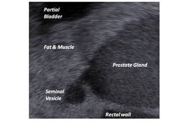
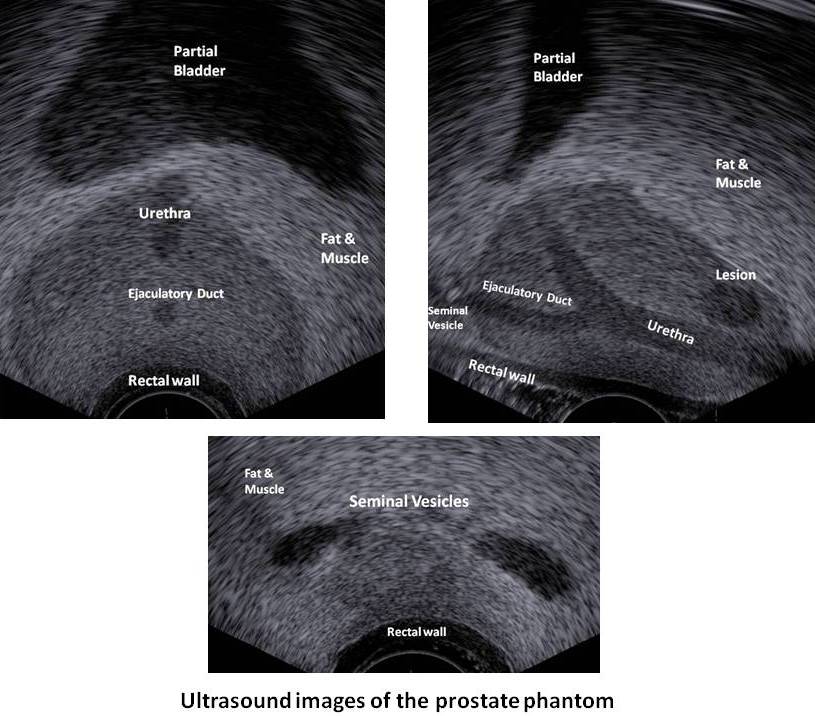
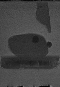
Ultrasound sagittal view of the prostate phantom
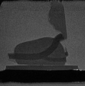
Sagittal MRI view of
the Prostate Phantom
Sagittal MRI view of
the Prostate Phantom
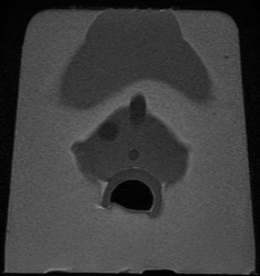
Prostate Phantom
MRI Transverse

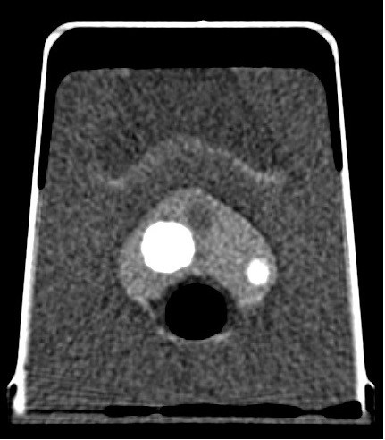
Prostate Phantom CT Transverse Scans
Enlarged Prostate Phantom CT
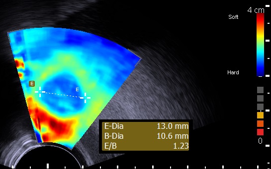
Prostate Phantom Elastography
Brachytherapy mode.
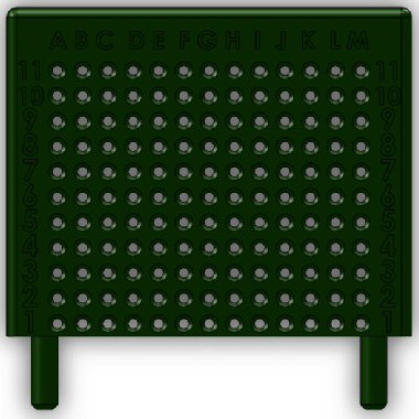
Brachytherapy template included with the model.
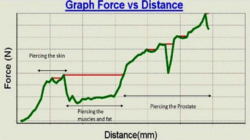
Real time graph (Prostate phantom device test bed) - of force as function of needle depth penetration.
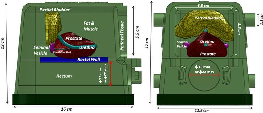
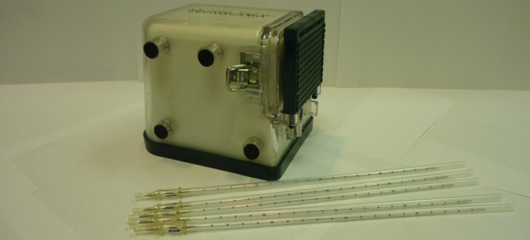
Internal tissues setup of the Brachytherapy prostate phantom. Diameter of 15 mm or 22 mm according to client demand.
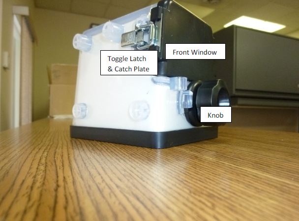
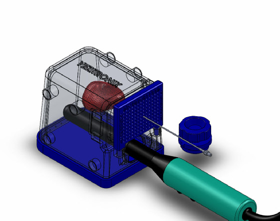
1.The Yezitronix Brachytherapy mode at its initial state when receiving the unit
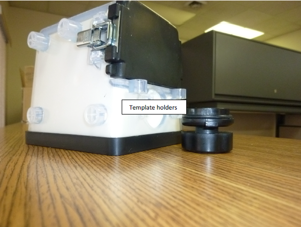
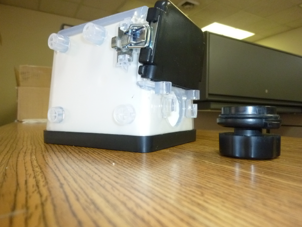
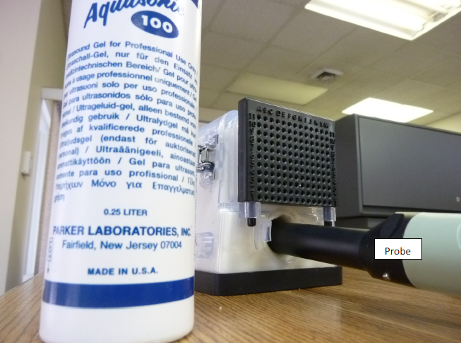
2.Remove the knob at the probe input channel by rotating it counterclockwise.
3.Release both (left and right) toggle latches from the catch plates in order to remove the front window.
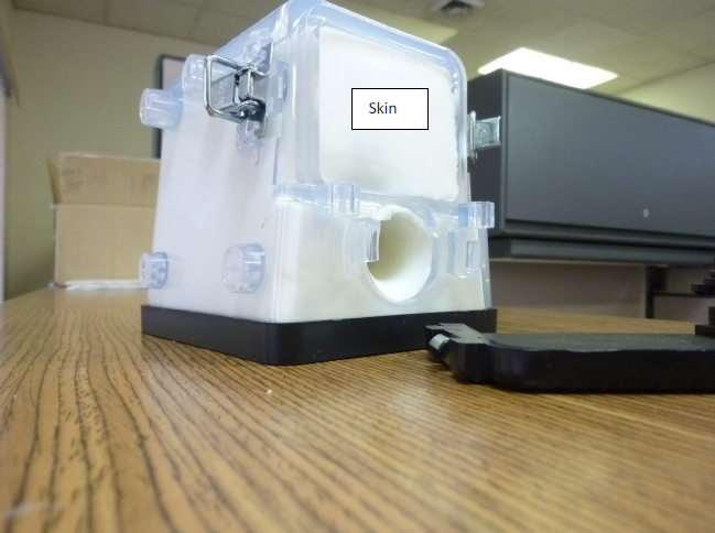
4.Remove the front window, to discover the skin component behind it.
5.Install the template Brachytherapy on the template holders.
6.Apply sufficient Ultrasound gel inside the probe channel before insertion of the Brachytherapy probe for better ultrasound image
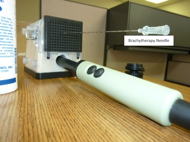
7.Insert the brachytherapy needle through the template holes and follow up its path using theultrasound planes (Transverse or Sagittal). Probe Brachytherapy Needle
8.When done with the experiments please go backward to:
a.Re-install the front window.
b.Remove the excess ultrasound gel from the probe channel.
c.Re-install the knob.
9.The steps in paragraph 8.,will keep your phantom fresh and ready for your next experiments.
Specifications :
Multi-layer material : each tissue or organ is independent and has its own characteristics defined by a real 3D shape, echogenicity level and mechanical properties.
There are 4 embedded lesions in the prostate to help simulate biopsies or brachytherapies procedures.
Multiple usage of the same packaged phantom over an extended period of time.
Enclosure :
16(L) x 11.5(W) x 12(H), Material . PVC, PC and metal latches. Front upper window 6.5(W) x 5.5 (H), Probe input diameter . 3.5 (all units in cm)
Perineal Tissue :
65(W) x 55 (H) x 3mm thick, approximate mechanical response of human tissue
Fat & Muscles :
Approximate mechanical response of human tissue
Urethra :
6mm diameter and 61mm(L)
Ejaculatory duct :
4mm diameter 28mm (L)
Seminal vesicles :
2 of 25(L)x6(W)x 4mm(Thick)
Prostate gland :
40cc, approximate mechanical response of human tissue.
Rectal wall :
81(L)x 75(W)x2.5(thick)mm, approximate mechanical response of human tissue.
Partial bladder :
13.4cc
Lesions :
4 Elliptic 0.3cc
Template:
13 columns and 11 rows.
ø: 2 mm
