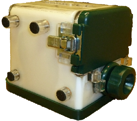
Prostate Phantom - Yezitronix Group Inc.
A left lateral decubitus position for TRUS (End fire biopsy mode)
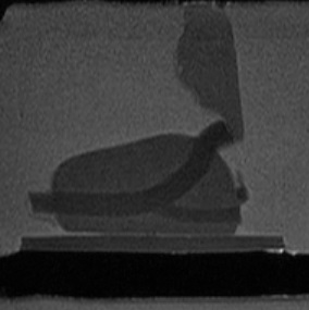
Sagittal MRI view of
the Prostate Phantom
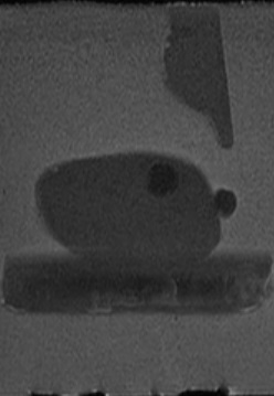
Sagittal MRI view of
the Prostate Phantom
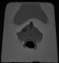
Prostate Phantom
MRI Transverse

Prostate Phantom CT Transverse Scans
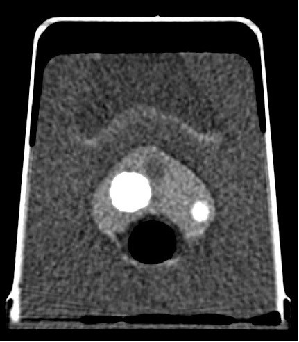
Enlarged Prostate Phantom CT
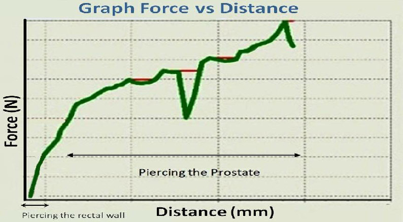
Real time graph (Prostate phantom device test bed) - of force as function of needle depth penetration
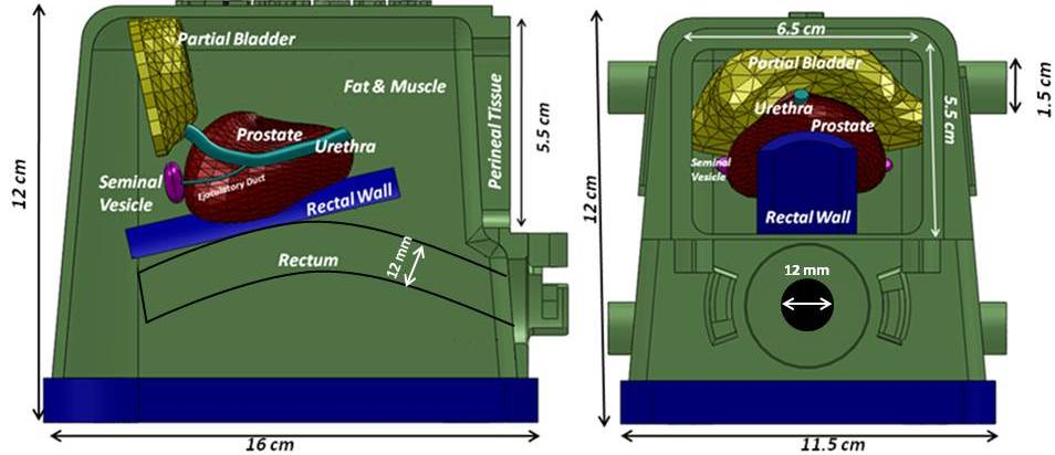
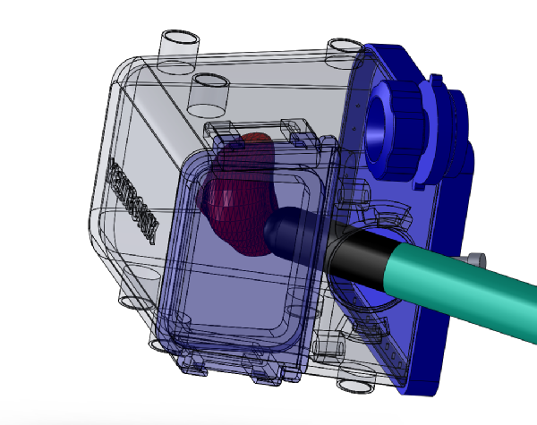
Internal tissues setup of the prostate phantom End fire model.
Specifications :
Multi-layer material : each tissue or organ is independent and has its own characteristics defined by a real 3D shape, echogenicity level and mechanical properties.
There are 4 embedded lesions in the prostate to help simulate biopsies or brachytherapies procedures.
Multiple usage of the same packaged phantom over an extended period of time.
Enclosure :
16(L) x 11.5(W) x 12(H), Material . PVC, PC and metal latches. Front upper window 6.5(W) x 5.5 (H), Probe input diameter . 3.5 (all units in cm)
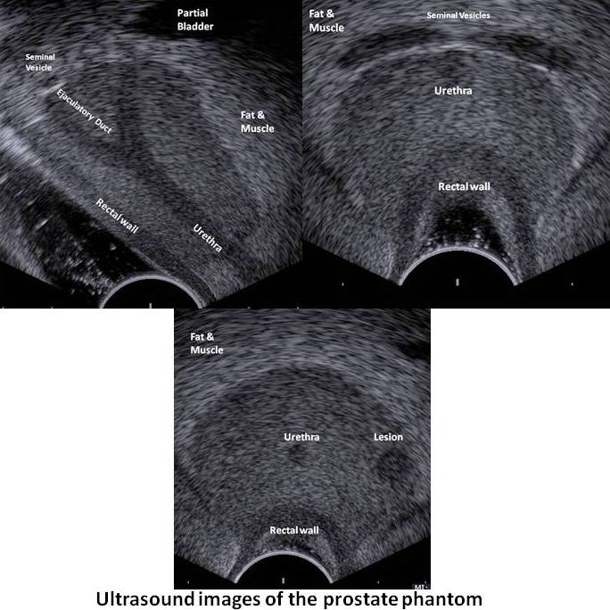
Perineal Tissue :
65(W) x 55 (H) x 3mm thick, approximate mechanical response of human tissue
Fat & Muscles :
Approximate mechanical response of human tissue
Urethra :
6mm diameter and 61mm(L)
Ejaculatory duct :
4mm diameter 28mm (L)
Seminal vesicles :
2 of 25(L)x6(W)x 4mm(Thick)
Prostate gland :
40cc, approximate mechanical response of human tissue.
Rectal wall :
81(L)x 75(W)x2.5(thick)mm, approximate mechanical response of human tissue.
Partial bladder :
13.4cc
Lesions :
4 Elliptic 0.3cc
Template:
13 columns and 11 rows.
ø: 2 mm