Multimodality Prostate Phantom

Most Accurate Prostate Phantom & Surrounding Tissues Mimicking Device for Ultrasound, MRI, CT modalities

Multimodality Prostate Phantom image png
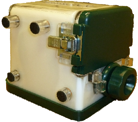
Yezitronix' Multi Modality Prostate Phantom Model S-MM-2.3 for Ultrasound, CT or MRI is a multiple usage phantom developed for training medical procedures such as ultrasound guided surgery, needle biopsy and radiation seed implantation.

Its ingenious and versatile design makes S-MM-2.3 model an incredible useful tool for training and simulation of different ultrasound rectal probes and surgical elements involves in prostate medical procedures.

The prostate phantom construct mimics the exact 3D shape size of:

40cc Prostate
Urethra
Seminal vesicle
Ejaculatory duct
Rectal wall
Partial bladder
Fat muscle tissues
Perineal Tissue
Numerous lesions located inside the prostate.

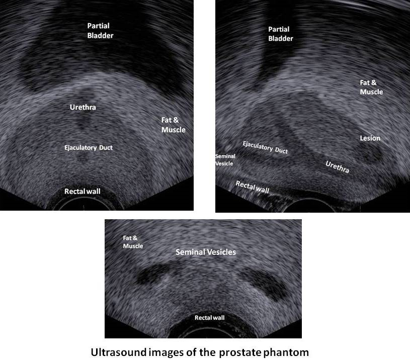
All organs & tissues are correctly adjusted to mimic exact ultrasound echogenicity seen by surgeon during OR procedures.
Suitable for:
OEMs. medical devices simulation, robotic system calibration, hospitals, clinics, med schools training and demonstrations.
Multimodality Prostate Phantom images png
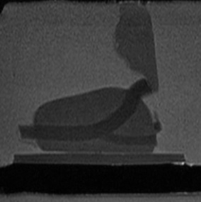
Sagittal MRI view of the Prostate Phantom 1
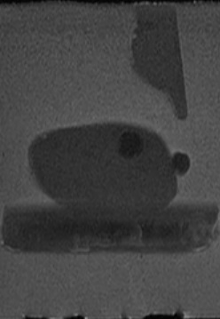
Sagittal MRI view of
the Prostate Phantom

Sagittal MRI view of the Prostate Phantom 2
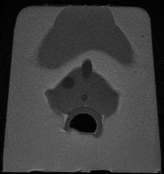
Sagittal MRI view of
the Prostate Phantom

MRI6.jpg

Prostate Phantom
MRI Transverse

Prostate Phantom CT Transverse Scans
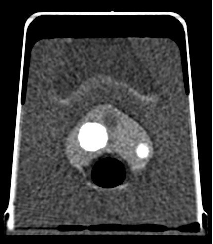
Prostate Phantom CT Transverse Scans

Enlarged Prostate Phantom CT
Enlarged Prostate Phantom CT

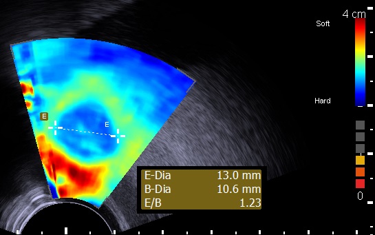
Prostate Elastography

Prostate Phantom Elastography

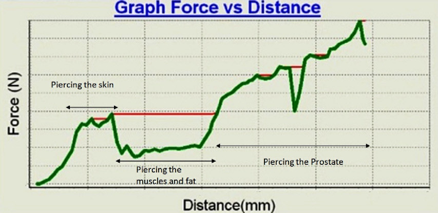
All phantom tissues' mechanical properties are approximated to human tissues. All prostate phantom layers mimic the tissue behaviour (dynamics) when pierced.

When for example a needle is piercing the Multi-Layer Prostate Phantom construct, it will generate a "Haptic Feedback (tactile)" to the user, simulating the real human tissue during biopsy or Brachytherapy procedure.

MultimodalityProstateP_graph.png

Real time graph (Prostate phantom device test bed) - of force as function of needle depth penetration
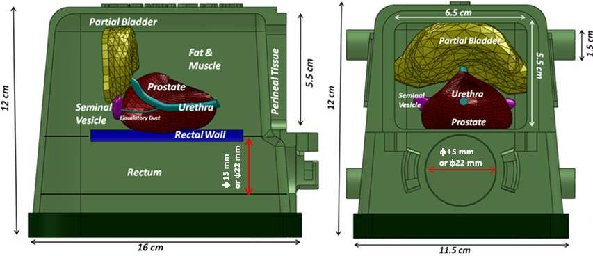
MultimodalityProstateP_schema.png

Internal tissues setup of the Multimodality prostate phantom


Specifications :

Multi-layer material : each tissue or organ is independent and has its own characteristics defined by a real 3D shape, echogenicity level and mechanical properties.
There are 4 embedded lesions in the prostate to help simulate biopsies or brachytherapies procedures.
Multiple usage of the same packaged phantom over an extended period of time.


Enclosure :

16(L) x 11.5(W) x 12(H), Material . PVC, PC and metal latches. Front upper window 6.5(W) x 5.5 (H), Probe input diameter . 3.5 (all units in cm)

Perineal Tissue :

65(W) x 55 (H) x 3mm thick, approximate mechanical response of human tissue

Fat & Muscles :

Approximate mechanical response of human tissue


Urethra :

6mm diameter and 61mm(L)

Ejaculatory duct :

4mm diameter 28mm (L)

Seminal vesicles :

2 of 25(L)x6(W)x 4mm(Thick)

Prostate gland :

40cc, approximate mechanical response of human tissue.


Rectal wall :

81(L)x 75(W)x2.5(thick)mm, approximate mechanical response of human tissue.

Partial bladder :

13.4cc

Lesions :

4 Elliptic 0.3cc

Template:

13 columns and 11 rows.
ø: 2 mm


Available Models :

  • Bi-plane (Side Fire) : S-BP-2.3

  • End fire mode : S-EF-2.3

  • Multi-modality mode (Ultrasound, CT, MRI) : S-MM-2.3

  • Without lesions : S-WL-2.3

  • Coloured components (Bladder, Urethra, Seminal Vesicles, Ejaculatory Duct) for Biopsy training : S-CC-2.3

  • With hollow Urethra for catheter insertion : S-HU-2.3

  • Custom design according to customer specifications : S-CD-2.3

Technology developed with the collaboration of the scientists from NRCC-National Research Council of Canada and surgeons at the department of urology at the London Health Sciences Centre in Ontario, Canada.
Patent Pending.
All components are based on Yezitronix' new unique MajesticMix material.
Speed of propagation ~1540m/s